How to interpret an echo report? (Please see the YouTube video above for illustrations) Echocardiogram, often called just echo in short is ultrasound imaging of the heart. Though
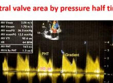
Mitral valve area by pressure half time Measurement of mitral valve area by pressure half time is an often applied method using Doppler echocardiography. Mitral valve area can
Segmental approach to congenital heart disease Segmental approach is used routinely in the echocardiographic evaluation of congenital heart disease. It is also useful in other cardiac imaging modalities
Echocardiographic assessment of patient-prosthesis mismatch Patient-prosthesis mismatch is present when the effective prosthetic valve area is less than that of the normal human valve [1]. The reduction in
Basic echocardiographic views Echocardiography is now not restricted to the echocardiographic laboratory. It is used in the emergency department, at bedside, in the intensive care unit as well
Annulus paradoxus and annulus reversus on tissue Doppler in constrictive pericarditis Annulus paradoxus The term “annulus paradoxus” was proposed by Ha JW et al to indicate the inverse
Estimation of PCWP from E/E’ on Tissue Doppler Imaging Conventionally, pulmonary capillary wedge pressure (PCWP) is measured using a catheter in the pulmonary artery. Pulmonary artery diastolic pressure
Elevated prosthetic valve gradients Elevated gradients across prosthetic valves can occur due to various reasons. But before declaring that prosthetic valve gradients are elevated, the usual gradient across
Low gradient severe mitral stenosis Low gradient severe mitral stenosis has been defined as mean transmitral gradient <10 mm Hg in patients with mitral valve area ≤1.5 cm2 [1].