Category: Echocardiography
Echocardiogram Library
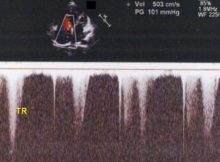
Echocardiogram in severe pulmonary hypertension with M-mode, 2D and Doppler recordings.
Read More
Echocardiography
Measurement of pressure half time in aortic regurgitation: CW tracing in apical 5C view showing measurement of pressure half time (P1/2t) in AR.
Read More
Echocardiography
AR jet with wrap around, seen because the velocity of the aortic regurgitation jet is more than the aliasing velocity setting.
Read More
Echocardiography
AR and MR - Colour Doppler echocardiogram: MR jet is seen in the LA in systolic frame on left side of image, while AR jet is seen in
Read More
Echocardiogram Library
M-mode echocardiogram in left ventricular dysfunction showing reduced contractile excursions of the interventricular septum (IVS) and left ventricular posterior wall (LVPW).
Read More
Echocardiography
Methods for assessing LV function by echo: Teicholz method
2. Simpson's two plane and three plane methods
3. Speckle tracking
4. 3D method
Read More
Echocardiogram Library
RHD, MR on colour Doppler echo shown in annotated and non annotated images.
Read More
Echocardiogram Library
TR Jet on CW Doppler: Continuous wave (CW) Doppler from apical 4C view across the tricuspid valve picked up this jet with peak gradient (PG) of 37 mm
Read More
Echocardiography
Myocardial contrast echocardiography (MCE) uses gas filled microbubbles to image the microcirculation of the heart.
Read More
Echocardiogram Library
Echocardiogram of RHD, MS, PSAX view: parasternal short axis view of rheumatic heart disease with mitral stenosis.
Read More
Posts navigation