Echo quiz – Cardiology MCQ The image is of: Echocardiogram in calcific mitral stenosis CT scan in aortic stenosis Bicuspid aortic valve on echocardiogram MR image of aortic
This parasternal short axis echocardiogram is a cross section of the left ventricle at the mitral valve level. Both anterior mitral leaflet (AML) and posterior mitral leaflet (PML)
Advantages of continuity equation for quantification of aortic stenosis Not affected by aortic regurgitation Can be used in the presence of left ventricular dysfunction, when gradient can be
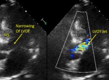
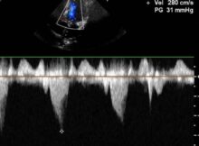
Echo quiz Discussion IVS (interventricular septum) is hypertrophied and bulges into the left ventricular outflow tract (LVOT), narrowing it. Gradient across the LVOT is increased to 26 mm
Mitral valve area by planimetry on echocardiogram Mitral valve area by planimetry on echocardiogram is usually obtained from the parasternal short axis view. It can also be obtained
LVOT gradient in HOCM – Doppler echocardiogram Left ventricular outflow tract gradient (LVOT) in hypertrophic obstructive cardiomyopathy (HOCM) is usually measured from the apical five chamber view (apical
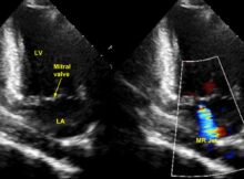
Mitral valve prolapse with regurgitation – echocardiogram Echocardiogram in apical four chamber view demonstrates prolapse of both anterior and posterior mitral leaflets. The central part of the leaflets
M-mode echocardiogram in left ventricular dysfunction M-mode echocardiogram is commonly used to measure left ventricular dimensions and ejection fraction. Ejection fraction is indicative of the left ventricular systolic
Mitral E/E’ for assessment of left ventricular diastolic function E/E’ measured using a combination of mitral flow Doppler and tissue Doppler of mitral annulus is an important measure
Doppler echocardiography in aortic stenosis AV Vmax: peak aortic velocity; AV Vmean: mean aortic velocity; AV maxPG: peak aortic gradient; AV mean PG: mean aortic gradient; AV VTI: