Parasternal long axis view in echocardiography
Parasternal long axis view in echocardiography
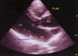
Parasternal long axis view is usually the first view which is obtained in echocardiography. The transducer is placed in the left parasternal region with the beam oriented to image the heart in the long axis view. RV: right ventricle; IVS: interventricular septum; AO: aorta; LV: left ventricle; LA: left atrium. The dense interface of the pericardium is seen posterior to left atrium and ventricle. But echocardiography is not that good for assessing the thickness of the pericardium, though it is excellent for assessment of pericardial effusion. This is because the interface is highly echogenic and the thickness can be overestimated as in this case.
Related Posts

About The Author
Johnson Francis
Former Professor of Cardiology, Calicut Govt. Medical Kozhikode, Kerala, India. Editor-in-Chief, BMH Medical Journal