Category: Echocardiogram Library
Echocardiogram Library
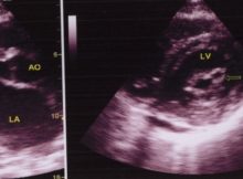
Echocardiogram in Mitral Stenosis Left panel shows the parasternal long axis view (PLAX). AO: Aorta; MVO: Mitral valve opening; LA: left atrium. Doming of the anterior mitral leaflet
Read More
Echocardiogram Library
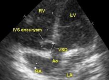
Color Doppler echocardiogram showing right to left shunt across a subaortic ventricular septal defect
Read More
Echocardiogram Library
Large subaortic VSD with aortic override - echocardiogram, seen from the parasternal long axis view
Read More
Echocardiogram Library
Trivial aortic regurgitation - echocardiogram seen on colour Doppler echocardiogram in the left image (AR)
Read More
Echocardiogram Library
Evaluation of prosthetic valves is one of the difficult tasks in echocardiography because of the dense shadows caused by prosthetic valves.
Read More
Echocardiogram Library
Ventricular septal defect: 2-D and Doppler echocardiograms
Read More
Echocardiogram Library
Vegetation on tricuspid valve seen on echocardiography from a modified apical four chamber view.
Read More
Echocardiogram Library
Parasternal long axis view is usually the first view which is obtained in echocardiography.
Read More
Echocardiogram Library
Echo showing right ventricle, aorta, left atrium, left ventricle and flail posterior mitral leaflet which points back into the left atrium during systole.
Read More
Echocardiogram Library
E/A reversal in Doppler echo tracing in left ventricular diastolic dysfunction.
Read More
Posts navigation