Pacing in complete heart block – ECG
Pacing in complete heart block – ECG
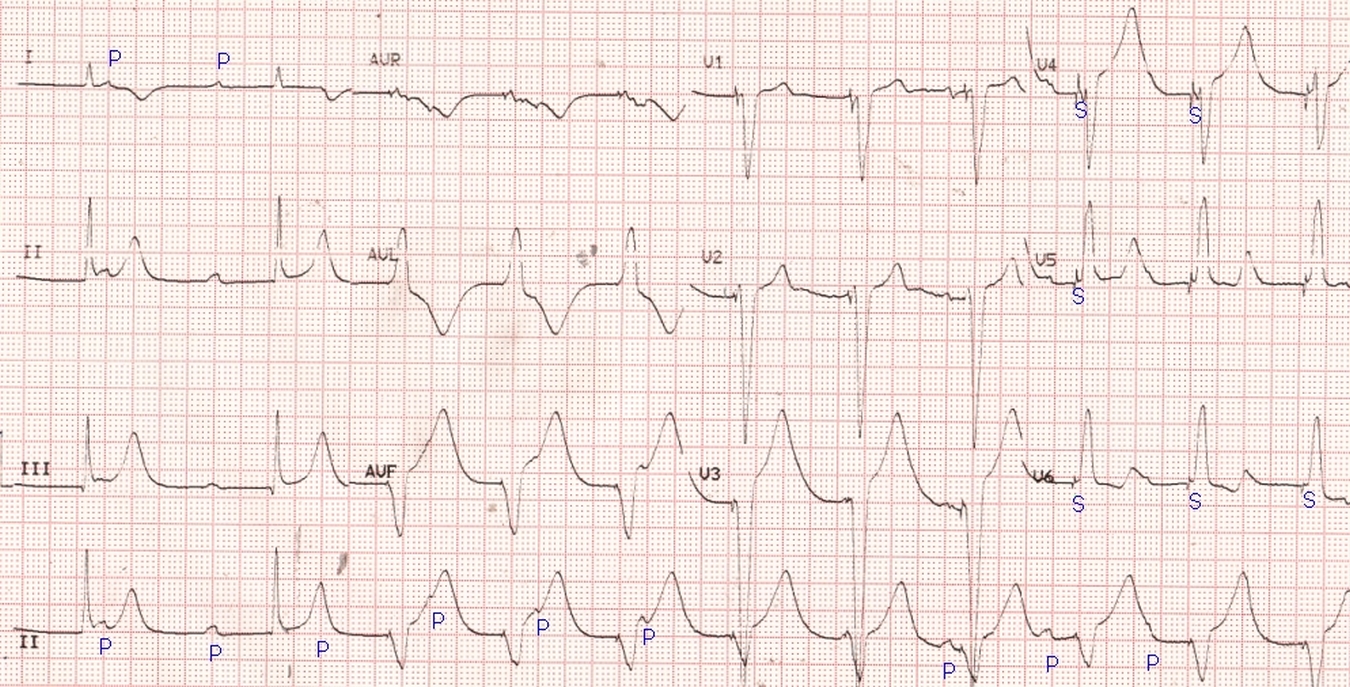
Pacing in complete heart block (CHB): Initial part of the ECG shows narrow QRS complexes at a rate of around 43/minute. Dissociated P waves can be seen at a rate of around 75/min. T wave inversion is noted in lead I. After the initial two complexes, the ECG shows a wide QRS rhythm at around 70/min, with a spike (S) seen just before the QRS complexes. In some leads the pacing spikes are quite small (aVF) or even not visible (aVL). Spike is well seen in most of the chest leads. Dissociated P waves are seen in the paced rhythm as well, as it is a ventricular pacing and not AV sequential pacing. Some of the P waves fall on the T wave and manifest as a notching of the T waves, but can be identified by their timing. In one case the P wave is at the apex of the T wave and makes the T wave peaked (T wave after the second QRS complex in the rhythm strip). Paced beats have a left bundle branch block pattern with negative QRS complexes in inferior leads, indicating pacing from the right ventricular apex. This pattern is seen as wide S or QS complexes in anterior leads and slurred R waves in lateral leads. T wave inversion in lateral leads in the paced beats is secondary T wave change due to primary abnormality of depolarization sequence, with right ventricle being activated first and left ventricle activated slowly by spread of impulse through the myocardium. QRS complexes are negative in inferior leads as the activation starts from the apical region and spreads upwards, away from the inferior leads.
Here is a short journal article on ECG in complete heart block, giving ECGs with and without pacing. There is right bundle branch block pattern as well.
Related Posts


About The Author
Johnson Francis
Former Professor of Cardiology, Calicut Govt. Medical Kozhikode, Kerala, India. Editor-in-Chief, BMH Medical Journal